The Devil Is In The Details: Predicting Alzheimer’s
The ability to predict the onset of Alzheimer’s disease is closer than you think
By Veronique Morin
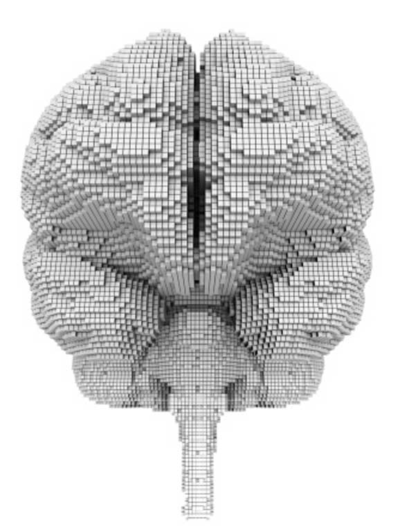
IN THE QUEST for early diagnosis, key to treating Alzheimer’s disease, scientists are now gazing into their crystal balls using something called a voxel. Voxels are units of volume – much like a pixel is a unit of a picture. But what on earth does this have to do with Alzheimer’s, you might ask? Well, by looking at the brain, voxel by voxel, or one cubic millimetre at a time, Alzheimer’s disease could be better diagnosed.
“The big focus right now is on prevention, to find new ways of identifying and treating the disease earlier, even before symptoms occur,” says Rosanne Meandro of the Alzheimer Society of Canada. The disease is a degenerative process that starts decades before symptoms become apparent. When they do, it might be too late for some treatments because certain parts of the brain, such as the hippocampus, which is responsible for memory, have been impaired. If Alzheimer’s disease is expected to more than triple by 2050 in Canada, the need to find proper diagnostic and predictive tools is pressing.
“The Holy Grail for doctors would be to assess someone in their 50s and predict whether that person will develop the disease 20 years down the road,” says Dr. Jens C. Pruessner, director of the McGill Centre for Studies in Aging and associate professor, department of psychiatry, McGill University. An expert in imaging of the aging brain, Pruessner was one of the co-authors on a 2012 research paper about SNIPE (scoring by nonlocal image patch estimator), an innovative computer method designed to assess and predict who will develop Alzheimer’s disease.
Now, what looks good on paper is closer to becoming an application. “What we found is revolutionary because it gives an accuracy never equalled before,” says Prof. Louis Collins, who led the team at the Montreal Neurological Institute of McGill University. An MR (magnetic resonance)image of the brain is divided into very tiny volumes (voxels) as small as one cubic millimetre, then, by comparing each tiny part with hundreds of other tiny parts of both healthy aging brains and brains of those who have developed Alzheimer’s disease, researchers have reached a diagnostic accuracy of 93 per cent.
“What is perhaps even more impressive,” says Collins, “is that this new tool can offer prognostic information for subjects with mild cognitive impairment (MCI) with 74 per cent accuracy.” Only about 10 per cent of people with mild cognitive impairment will develop Alzheimer’s disease.
With current methods, it is not definitive who will or who will not.
But Collins says that with this powerful statistical method, his team has been able to identify very subtle changes in the brain that are undetectable using current methods.
RELATED POST: Walk Your Way to Better Brain Health
“We are able to detect changes in the morphology of the hippocampus that we can now associate with the early development of Alzheimer’s disease,” explains Dr. Pierrick Coupé, the main author of the research and a computer scientist with the Laboratoire Bordelais de Recherche en Informatique in Bordeaux, France, who chose to work with the Montreal Neurological Institute because of its reputation. Coupé describes the process thus: “When we compare two pictures, we naturally try to find correspondences between a restricted number of subparts in both images. The evaluation of all the possible matching between all the image subparts is too complex. Now, imagine that you want to compare 100 images at the same time based on this exhaustive search … By programming a computer to achieve this task, researchers can detect subtle signs of Alzheimer’s disease in your brain where the human eye would not be able!”
“The jury is still out as we will be waiting for replication of these results,” says Yves Joanette, scientific director of the Institute of Aging of the Canadian Institute for Health Research (CIHR), the main funding agency in Canada. “But this is an example of the strength of Canadian neuroimaging research.”
ALSO ON EVERYTHINGZOOMER:
The Zoomer Report: Driving With Dementia
The Brainiac: Aging, Sex & Memory
Alzheimer’s disease: Signs to Watch For
'%3E %3Cg id='Group'%3E %3Cpath id='Vector_2' d='M12.4876 13.8996V13.4213H0.743091V15.2149H9.18805L9.22196 15.2685C9.19653 15.2272 3.24373 25.2836 0.74521 29.5469C0.420975 30.1015 0.0797856 30.7158 0.0797856 30.7158C0.0713089 30.7323 0.0585938 30.755 0.0585938 30.755H12.1401V28.9614H3.67181L3.6379 28.9078C3.37512 29.1222 12.4876 13.8996 12.4876 13.8996ZM36.1229 22.115C36.1229 26.4896 33.7537 29.18 31.1683 29.18C28.5829 29.18 26.1861 26.4896 26.1861 22.115C26.1861 17.7403 28.5553 15.0231 31.1683 15.0231C33.7812 15.0231 36.1229 17.7403 36.1229 22.115ZM22.0155 22.115C22.0155 26.4896 19.6462 29.18 17.0587 29.18C14.4712 29.18 12.0765 26.4896 12.0765 22.115C12.0765 17.7403 14.4458 15.0231 17.0587 15.0231C19.6717 15.0231 22.0155 17.7403 22.0155 22.115ZM38.223 22.0613C38.223 16.7095 35.0443 13.1492 31.1661 13.1492C27.288 13.1492 24.3402 16.4312 24.0987 21.4326C23.8592 16.4333 20.7842 13.1492 17.0587 13.1492C13.3332 13.1492 9.97427 16.7074 9.97427 22.0613C9.97427 27.4153 13.1785 31.0003 17.0587 31.0003C20.9389 31.0003 23.8592 27.7183 24.0987 22.6922C24.3402 27.7162 27.4406 31.0003 31.1661 31.0003C34.8917 31.0003 38.223 27.4421 38.223 22.0613ZM51.3747 30.755H53.5257L52.3178 13.4213H50.1668L45.797 27.3328L41.4273 13.4213H39.2763L38.0662 30.755H40.2172L41.0352 19.0247L44.7205 30.755H46.8714L50.5567 19.0247L51.3747 30.755ZM53.9453 30.755H62.3797V28.9614H56.0963V22.9293H60.964V21.1357H56.0963V15.2128H61.8859V13.4192H53.9453V30.7529V30.755ZM69.3624 18.2021C69.3624 20.1029 68.1333 21.1625 66.7367 21.1625H64.893V15.2128H66.7092C68.1333 15.2128 69.3624 16.3261 69.3624 18.2021ZM71.5134 18.1753C71.5134 15.2128 69.3348 13.4213 67.0164 13.4213H62.7993V30.755H64.893V22.9313H66.7092L71.5112 30.755H73.997L68.8601 22.4695C70.369 21.8717 71.5134 20.3502 71.5134 18.1773' fill='%23231F20'/%3E %3C/g%3E %3C/g%3E %3C/g%3E %3Cg id='Group_2'%3E %3Cpath id='Vector_3' d='M9.67285 8.56629V3.50513H12.6249V4.11535H10.5947V5.71719H12.2943V6.32742H10.5947V7.954H12.9046V8.56423H9.67285V8.56629Z' fill='%23231F20'/%3E %3Cpath id='Vector_4' d='M17.6819 8.6137H17.2581L15.1855 3.50513H16.1392L17.0822 5.86975C17.2178 6.20991 17.362 6.63872 17.4976 7.05103H17.5188C17.6544 6.64696 17.7879 6.2264 17.9341 5.86975L18.8666 3.50513H19.7482L17.6862 8.6137H17.6819Z' fill='%23231F20'/%3E %3Cpath id='Vector_5' d='M22.4072 8.56629V3.50513H25.3593V4.11535H23.3291V5.71719H25.0287V6.32742H23.3291V7.954H25.639V8.56423H22.4072V8.56629Z' fill='%23231F20'/%3E %3Cpath id='Vector_6' d='M31.6164 8.56629L29.9698 6.32123H29.2959V8.56629H28.374V3.50513H30.0948C30.9552 3.50513 31.805 4.01227 31.805 4.90906C31.805 5.54402 31.3896 5.97283 30.8725 6.16249L32.6654 8.56629H31.6185H31.6164ZM29.9592 4.10711H29.2959V5.71719H29.9592C30.436 5.71719 30.8704 5.43888 30.8704 4.90906C30.8704 4.37923 30.436 4.10711 29.9592 4.10711Z' fill='%23231F20'/%3E %3Cpath id='Vector_7' d='M37.5849 6.27382V8.56629H36.6737V6.28206L34.6943 3.50513H35.667L36.6503 4.90081C36.8156 5.13789 37.0021 5.38528 37.1378 5.59968H37.159C37.3031 5.39353 37.5001 5.11522 37.657 4.90081L38.6403 3.50513H39.5621L37.5828 6.27382H37.5849Z' fill='%23231F20'/%3E %3Cpath id='Vector_8' d='M44.3084 4.11535V8.56629H43.376V4.11535H41.7188V3.50513H45.9656V4.11535H44.3084Z' fill='%23231F20'/%3E %3Cpath id='Vector_9' d='M52.0484 8.56629V6.32948H49.499V8.56629H48.5771V3.50513H49.499V5.71926H52.0484V3.50513H52.9808V8.56629H52.0484Z' fill='%23231F20'/%3E %3Cpath id='Vector_10' d='M56.2129 8.56629V3.50513H57.1453V8.56629H56.2129Z' fill='%23231F20'/%3E %3Cpath id='Vector_11' d='M64.7402 8.64463L62.4091 6.17693C62.0467 5.79553 61.6208 5.31106 61.2796 4.91524L61.2584 4.92349C61.2796 5.3523 61.2902 5.7811 61.2902 6.13776V8.56629H60.3789V3.50513H61.0316L63.1868 5.82233C63.4878 6.14806 63.9222 6.63872 64.2337 7.00362L64.2549 6.99537C64.2337 6.62223 64.2231 6.17899 64.2231 5.83677V3.50513H65.1344V8.64669H64.7402V8.64463Z' fill='%23231F20'/%3E %3Cpath id='Vector_12' d='M70.9048 8.64416C69.4235 8.64416 68.0566 7.69172 68.0566 6.03421C68.0566 4.37671 69.4659 3.41602 70.8752 3.41602C71.5999 3.41602 72.17 3.57476 72.5324 3.78091L72.3883 4.42412C72.0471 4.21797 71.5491 4.05098 71.0002 4.05098C69.9639 4.05098 68.9912 4.77253 68.9912 6.04246C68.9912 7.31239 69.9448 8.01745 70.9599 8.01745C71.405 8.01745 71.7567 7.93911 71.975 7.7948V6.61352H70.8349V6.03421H72.8354V8.12877C72.3586 8.47718 71.7271 8.64416 70.9091 8.64416H70.9048Z' fill='%23231F20'/%3E %3C/g%3E %3Cpath id='Vector_13' d='M0.0839844 1.00212L73.997 1' stroke='%23231F20' stroke-width='1.25' stroke-miterlimit='10'/%3E %3Cpath id='Vector_14' d='M0.0839844 10.9116H73.997' stroke='%23231F20' stroke-width='1.25' stroke-miterlimit='10'/%3E %3Cpath id='Vector_15' d='M4.91016 4.15649L3.55812 4.13794L5.18777 5.7068L0 5.6532V6.5974L5.20049 6.651L3.61321 8.18068L4.96526 8.19718L7.0018 6.20157L4.91016 4.15649Z' fill='%23D71920'/%3E %3C/g%3E %3C/svg%3E)
